Veterinary Blog - Knee-d to Know: Medial Patellar Luxation Treatment Strategies | Movora
Learn about the latest surgical options to improve your patient’s quality of life after a diagnosis of MPL.
Medial patellar luxation causes lameness, pain, and arthritis. Surgical intervention helps reset stifle anatomy, alleviating further damage mitigating pain. From femoral trochleoplasty to patellar groove replacement to correcting femoral deformities, Movora offers tools to help. Movora Product Design Engineer Christina Rohlf, who earned a Ph.D. in Biomedical Engineering from the University of California, Davis, explains Movora’s expansive portfolio of surgical options for MPL surgery.
Veterinarians frequently diagnose medial patellar luxation (MPL), a pathology of the canine stifle that causes hindlimb lameness, osteoarthritis, and pain. MPL ranks seventh among canine orthopedic diagnoses in the UK.1 The prevalence of MPL in the US is 5.4%.1
While MPL frequently afflicts small breed dogs like Pomeranians, Chihuahuas, French Bulldogs, and Yorkshire Terriers, it also affects large breed dogs.2
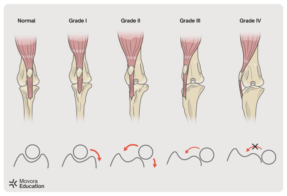
Clinical signs determine MPL severity. Veterinarians use a grading scale based on symptoms to classify MPL.3
- Grade I: The patella rarely luxates spontaneously but will luxate with pressure during a physical exam. It reduces spontaneously.
- Grade II: Lateral pressure or manual extension and flexion of the stifle displaces the patella. Femoral deformities may exist.
- Grade III: The patella is luxated almost all the time. Manual reduction of the patella is possible. The patella will reluxate with stifle flexion and extension. Femoral, tibial, and soft tissue abnormalities are present.
- Grade IV: The patella is permanently luxated. Manual pressure cannot reduce the patella. Other soft tissue and bone abnormalities are notable.

Treatment Options
MPL treatment includes conservative care or surgery.3 Treatment choice depends on the patient’s clinical background, physical examination findings, luxation frequency, and age. 3
Veterinarians generally monitor Grade I MPL patients for symptoms. Experts recommend surgery for all MPL patients with lameness, especially young patients who risk growth plate abnormalities and rapid cartilage degradation. Grade III and IV MPL typically require surgery to prevent articular cartilage damage and osteoarthritis.4
Femoral Trochleoplasty
Femoral trochleoplasty, a standard treatment for MPL, deepens the trochlear groove to prevent patellar luxation.
Movora offers the novel NGD Semi-Cylindrical Recession Trochleoplasty (SCRT) system. It precisely controls trochleoplasty depth with a rounded trochleoplasty cut and polymer cutting guides. Compared to other trochleoplasty techniques, the SCRT system reduces the chance of fracturing the trochlear ridges, increases the depth of the patella in the trochlear groove, and eliminates the gap between the resected and native bone.5 The SCRT instrumentation creates accurate cuts in 1.5mm increments to treat small dogs and cats consistently and safely. 6
Patellar Groove Replacement
Surgeons cannot perform femoral trochleoplasty in cases of cartilage erosion, malformation, or a prior trochleoplasty procedure requiring revision.
Movora offers a patellar groove replacement (PGR) device to address these situations. The KYON PGR has two components:
- A porous base plate that promotes bone ingrowth
- A trochlear groove coated with amorphous, diamond-like carbon that creates a low friction, wear-resistant bearing surface for the patella
In a study of PGR in 35 dogs with grade II to IV patellar luxation, 24 dogs had complete resolution of lameness 12 weeks post-op, and 31 dogs showed no lameness at long-term follow-up.7
Multifactorial Treatment of MPL
MPL, often a multifactorial condition, may require more invasive treatments for complete resolution. Movora has solutions.
Distal Femoral Deformities
Studies show that distal femoral deformities accompany severe MPL.8 Targeted osteotomy to correct these femoral deformities promotes patellar reduction when other treatments fail.3
- Movora offers implants to stabilize the bone following a corrective osteotomy: A pre-contoured distal femoral osteotomy plate stabilizes a lateral closing wedge osteotomy
- The I-Loc Interlocking Nail stabilizes a medial opening wedge osteotomy
Concurrent CrCL Rupture
Studies show up to 34% of MPL cases have concurrent CrCL rupture.9 Movora offers modified tibial tuberosity advancement (TTA) and tibial plateau leveling osteotomy (TPLO) implants for surgeons treating concurrent CrCL rupture.
Modified TTA Technique
To modify the TTA technique, the surgeon places a spacer under the cranial wing of the TTA cage, allowing for lateral transposition of the tibial tuberosity and advancement. In a study of this technique, 35 out of 39 dogs with grade II to IV MPL had no lameness at long-term follow-up.10
Modified TPLO Technique
To modify the TPLO technique, Movora offers the Offset Evolution TPLO plate in October 2023. This plate features a pre-contoured sharp bend to provide medial translation of the tibial plateau. Sixteen Offset Evolution TPLO plates are available to accommodate each patient’s desired plate size and offset distance.
Movora: Your Complete Source for MPL Treatment Solutions
Whether you need a better trochleoplasty procedure or surgical implants to address complex MPL cases, Movora has solutions. Our comprehensive product portfolio, backed by years of research and development, helps you unleash optimal results. Consult with your regional sales representative to discuss how our MPL treatment options can help you deliver superior patient care and to find the right technique for your practice. Or browse the options for yourself at education.movora.com for introductory and advanced courses from Movora Education.
References
1. Perry KL, Déjardin LM. Canine medial patellar luxation. J Small Anim Pract. 2021;62(5):315-335.
2. O’Neill DG, Meeson RL, Sheridan A, Church DB, Brodbelt DC. The epidemiology of patellar luxation in dogs attending primary-care veterinary practices in England. Canine Genet Epidemiol. 2016;3(4).
3. Fossum TW. Small Animal Surgery. 5th ed. (Elsevier, Inc, ed.). Mosby; 2019.
4. Patellar luxations. American College of Veterinary Surgeons. Published June 16, 2023. Accessed May 13, 2024. https://www.acvs.org/small-animal/patellar-luxations/
5. Blackford-Winders CL, Daubert M, Rendahl AK, Conzemius MG. Comparison of Semi-Cylindrical Recession Trochleoplasty and Trochlear Block Recession for the Treatment of Canine Medial Patellar Luxation: A Pilot Study. Vet Comp Orthop Traumatol. 2021;34(3):183-190.
6. Deom K, Conzemius MG, Tarricone J, Nye C, Veytsman S. Short term outcome for surgical correction of feline medial patellar luxations via semi cylindrical recession trochleoplasty. JFMS Open Rep. 2023;9(2).
7. Dokic Z, Lorinson D, Weigel JP, Vezzoni A. Patellar groove replacement in patellar luxation with severe femoro-patellar osteoarthritis. Vet Comp Orthop Traumatol. 2015;28(2):142-130.
8. Yasukawa S, Edamura K, Tanegashima K, et al. Evaluation of bone deformities of the femur, tibia, and patella in Toy Poodles with medial patellar luxation using computed tomography. Vet Comp Orthop Traumatol. 2016;29(1):29-38.
9. Campbell CA, Horstman CL, Mason DR, Evans RB. Severity of patellar luxation and frequency of concomitant cranial cruciat e ligament rupture in dogs 162 cases. J Am Vet Med Assoc. 2010;236(8):887-891.
10. Yeadon R, Fitzpatrick N, Kowaleski MP. Tibial tuberosity transposition-advancement for treatment of medial patellar luxation and concomitant cranial cruciate ligament disease in the dog. Surgical technique, radiographic and clinical outcomes. Vet Comp Orthop Traumatol. 2011;24(1):18-26.
